Scanning Near-field Optical Microscopy (SNOM)
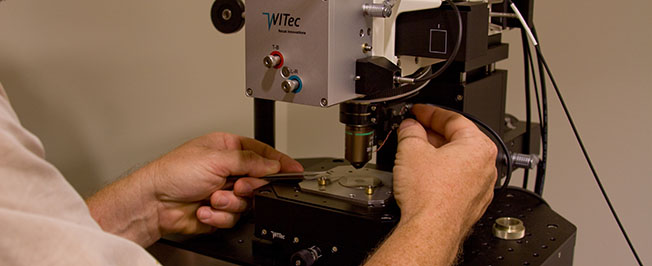
WITEC alpha300 S Scanning Near-field Optical Microscope

Schematic: The excitation laser light is focused through an aperture with a diameter smaller than the excitation wavelength, resulting in a near field on the far side of the aperture.
Instrument Calendar:
SNOM features:
- Micro-fabricated cantilever SNOM sensors
- Aperture size typically 100 nm, other optional
- Standard AFM cantilever probes
- Beam-deflection distance control for SNOM and AFM
- Low noise, highly focused optics for beam deflection laser
- No interference with excitation laser
- Ultra-low laser noise
- Near-field Mode 100 nm, depending on aperture size; Confocal Mode typically 200 nm diffraction-limited
Typical Applications:
- As scanning near-field microscopy requires only minimal sample preparation if any, it is ideally suited to quickly and effortlessly image the optical properties of a sample with resolution below the diffraction limit.
- Typical applications are found in nanotechnology research and particularly in the highly relevant fields of Nano-Photonics and Nano-Optics.
- In Life Science and materials research, SNOM allows the optical detection of the most miniscule surface structures of transparent as well as opaque samples.
- Using fluorescence techniques, even single molecule detection is easily achievable.

Switching from confocal to near-field allows portions of a sample to be measured with high resolution, like this 12 μm x 12 μm scan of a single human leukocyte in transmission.

Confocal fluorescence image of DNA strands solved in liquid, marked with ethidium bromide, then excited with an argon ion laser.